
Chronic Subdural Hematoma
Chronic subdural hematoma (CSDH) is a common neurological condition characterized by the slow accumulation of blood in the space between the brain's surface and its outermost covering (the dura mater). Unlike acute subdural hematomas, which develop rapidly after severe head trauma, chronic ones often develop gradually over weeks to months and are more prevalent in older adults.
Causes and Risk Factors
CSDH typically results from minor head trauma that tears fragile bridging veins between the brain and dura. In many cases, patients may not recall any injury. Key risk factors include:
- Advanced age (brain atrophy increases the space where blood can collect)
- Use of anticoagulant or antiplatelet medications (e.g., warfarin, aspirin)
- Alcohol abuse or liver disease
- History of falls or previous brain injury
- Conditions causing brain shrinkage, such as dementia or prior stroke
The condition is more common in men and those over age 60. Bilateral (both sides) hematomas can also occur.
Symptoms
Symptoms of chronic subdural hematoma develop insidiously and can mimic other conditions like dementia or stroke. Common signs include:
- Persistent or fluctuating headache
- Confusion, memory problems, or personality changes
- Weakness or numbness on one side of the body
- Difficulty walking or balance issues
- Drowsiness, seizures, or in severe cases, coma
Because symptoms progress slowly, diagnosis is sometimes delayed.
Diagnosis
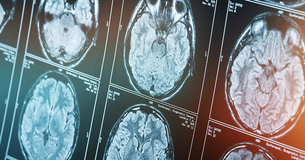
Imaging is essential for diagnosis. A non-contrast CT scan of the head is the preferred initial test, revealing a crescent-shaped collection of blood. MRI may provide additional detail. Neurological examination helps assess severity. Doctors also review medication history and coagulation status.
Treatment Options
Management depends on the size of the hematoma, symptom severity, and patient overall health.
- Observation: Small, asymptomatic CSDHs may resolve spontaneously with close monitoring and serial imaging.
- Medications: Corticosteroids, tranexamic acid, or other agents are sometimes used to reduce inflammation or promote resolution, though evidence varies.
- Surgical Drainage: The mainstay for symptomatic or large hematomas. Procedures include burr hole drainage (small holes drilled in the skull to evacuate the fluid) or twist-drill craniostomy. These are often performed under local anesthesia and have high success rates. A subdural drain may be left temporarily to prevent recurrence.
Recurrence occurs in 10–20% of surgical cases, sometimes requiring repeat intervention.
Prognosis and Recovery
With prompt treatment, most patients experience good recovery, though older adults or those with significant comorbidities may have lingering neurological deficits. Rehabilitation with physical, occupational, and speech therapy can aid recovery. Prevention focuses on fall risk reduction, careful medication management, and protective headgear when appropriate.
Conclusion
Chronic subdural hematoma is a treatable condition, but early recognition is crucial to prevent complications. Advances in minimally invasive techniques and medical therapies continue to improve outcomes. Anyone experiencing unexplained neurological changes, especially after minor head injury or while on blood thinners, should seek prompt medical evaluation.
This article is for informational purposes only and does not replace professional medical advice. Consult a healthcare provider for any health concerns.